
Image Guided Endoscopic Sinus Surgery
Endoscopic sinus surgery (ESS) is a minimally invasive outpatient procedure used in the treatment of many nasal and sinus conditions. In ESS, small and narrow instruments are placed in the nostrils and passed through the nasal and sinus cavities with the aid of a rigid endoscope. The endoscope provides the surgeon with a magnified, but distorted, image of the surrounding diseased and normal structures. The instruments are used to remove diseased sinus tissue or open obstructed areas of the nose and sinuses.
Safety during sinus surgery cannot be over emphasized. The anatomic relationship of the nasal and sinus passages to the brain and eye creates the potential for significant, albeit rare, complications. Image-guided endoscopic sinus surgery allows for highly accurate visual updates during your sinus surgery. In IGS, your CT scan images are digitally linked to a computerized image-guidance system displayed on an operating room monitor via a sophisticated software program. Your CT scans are coupled to the surgical instruments by an electromagnetic tracking system. This gives the surgeon the ability to navigate, in real-time, the surgical instruments within the diseased sinuses, and correlate their position with vital structures while watching the monitor.
IGS requires a special image-guidance protocol technique when the preoperative computed tomography (CT) scan of the sinuses is performed. This may require additional imaging if a previous CT scan was performed without this special protocol.
Current evidence supports the use of this technology in specific instances for reducing intraoperative complications, and therefore, maximizing theoutcome of your surgery.1-3 Both pediatric and adults benefit from this technology.4 This technology is particularly useful when a patient has polyps or needs revision sinus surgery in which case normal anatomic structures are distorted. Not all sinus surgeries require image guidance. Feel free to ask your surgeon about this technology and if your surgery requires this technology. The Academy of Otolaryngology has a list of indications for using IGS during sinus surgery and other useful information for sinus surgery.
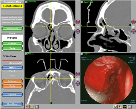
Surgeon's view of the computer monitor showing the sagittal (top left), axial (bottom left), and coronal (top left) images in real-time during the surgery. The fourth window (bottom right) is the real time operating video. The location at the center of the 3 CT images represents the tip of the surgical instrument seen on the video image. This case demonstrates revision sinus surgery in the right frontal sinus.
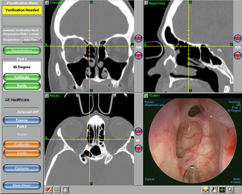
Surgeon’s view of the right skull base (top of the ethmoid sinus) demonstrating an important vascular structure, the anterior ethmoid artery. The coronal image (top left) confirms this anatomic structure noted by the indentation of the bone of the orbit (eye) seen just above and left of the cross hairs.